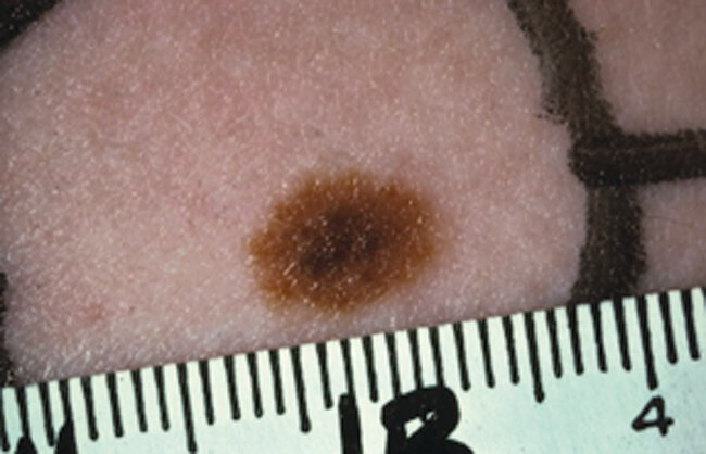
Often asymmetric, irregularly bordered, have more than one shade of brown, and may be quite large in diameter.
Many have a “fried egg” appearance, with an eccentric dark brown papular center and surrounding light brown ring
Dysplastic nevi can display one or more of the identifying characteristics of melanoma (asymmetry, irregular border, multiple colors, diameter >6 mm, evolution over time),
Pathophysiology :
Diagnostic Testing:
Requires excisional skin biopsy to remove the entire lesion should be performed for diagnosis. Very similar to melanoma in appearance, making an accurate clinical diagnosis impossible.
Treatment :
Prognosis:
Although the potential for an individual dysplastic nevus to develop into melanoma is low, patients with dysplastic nevi are at increased risk for developing a melanoma.
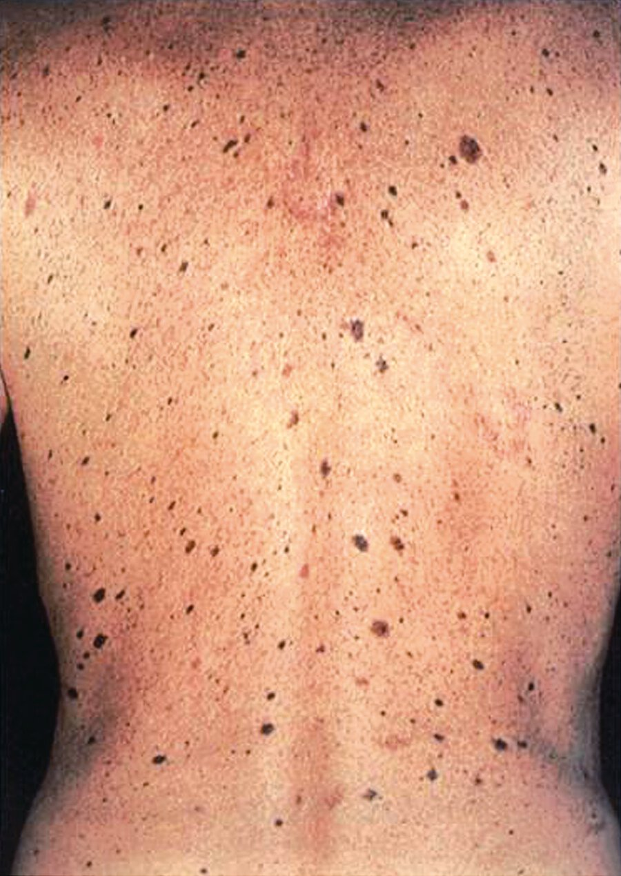
Patients with multiple dysplastic nevi are also at risk for developing melanoma and should be monitored closely
Some of these patients have Dysplastic Nevus Syndrome. Criteria for this syndrome include a history of melanoma in one or more first- or second-degree relatives; the presence of a large number of nevi (>50), as shown; multiple nevi having atypical clinical features; and multiple nevi that have atypical histologic features. These patients are at increased risk for melanoma and should have yearly full-skin examinations.